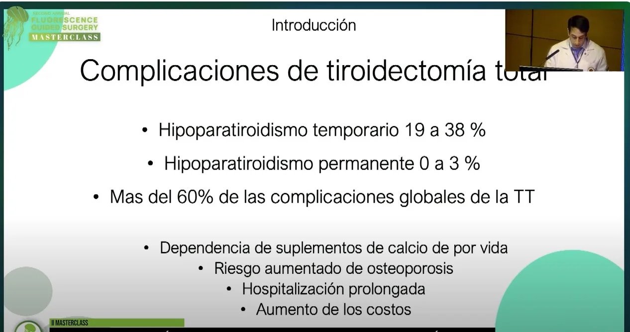
Título del video: Intraoperative Parathyroid Gland Management by Early and Advanced Career Surgeons Based on Viability Assessment by Visual Perception and Indocyanine Green Fluorescence Imaging
Título del video: ICG guided Robotic Adrenalectomy
Speakers: Dogukan Akkus, MD
Descripción: This video demonstrates a robotic adrenalectomy enhanced with indocyanine green (ICG) fluorescence imaging. The technique improves visualization of vascular structures and helps delineate adrenal tissue in real time, supporting a safer and more precise resection.
This video demonstrates a robotic adrenalectomy enhanced with indocyanine green (ICG) fluorescence imaging. The technique improves visualization of vascular structures and helps delineate adrenal tissue in real time, supporting a safer and more precise resection.
Indocyanine green (ICG) fluorescence imaging is widely used in lower limb lymphedema to visualize lymphatic flow and assess the severity of lymphatic dysfunction. This technique enables real-time mapping of superficial lymphatic vessels, supporting accurate diagnosis, staging, and surgical planning.
ICG lymphangiography has demonstrated high sensitivity and specificity compared to conventional imaging, allowing precise identification of functional lymphatic channels and guiding procedures such as lymphaticovenular anastomosis.
In the EMEA region, standardized dosing and timing protocols help ensure consistent fluorescence imaging and improve intraoperative decision-making in the management of lower limb lymphedema.
Sección:
Tabla:
Columna1:
Region of interest
, Contenido:
Lymphatic vessels
Columna1:
Dose
, Contenido:
0.05–0.1 mL (0.125-0.25 mg/mL)
Columna1:
Route
, Contenido:
Intra-dermal
Columna1:
Injection time
, Contenido:
Beginning of assessment
Columna1:
First ICG detection
, Contenido:
Beginning of assessment
Columna1:
ICG Duration
, Contenido:
Lymph flow tracked for 1 hr
Columna1:
Camera requirements
, Contenido:
NIR light source camera
Columna1:
References (full ref details at end of this guide doc)
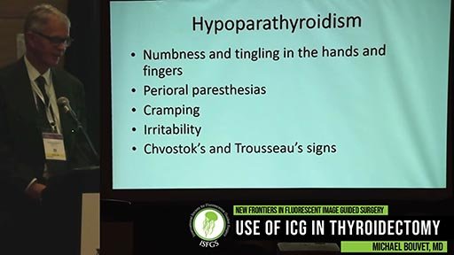
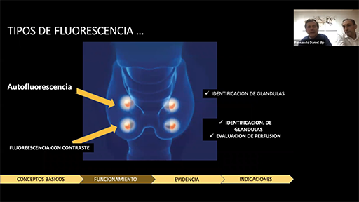
Indocyanine green (ICG) fluorescence imaging is used in thyroidectomy to identify and assess the perfusion of parathyroid glands, helping to preserve their function during surgery. Real-time visualization of vascularization supports intraoperative decision-making and reduces the risk of postoperative complications such as hypocalcemia and hypoparathyroidism.
In the EMEA region, standardized dosing and timing protocols aim to improve consistency in fluorescence imaging and enhance surgical safety by optimizing parathyroid preservation.
Sección:
Tabla:
Columna1:
Region of interest
, Contenido:
Parathyroid glands preservation - anatomy of feeder vessels to Parathyroid glands
Columna1:
Dose
, Contenido:
0.1mg/kg
Columna1:
Route
, Contenido:
I.V.*
Columna1:
Injection time
, Contenido:
Intraoperatively
Columna1:
First ICG detection
, Contenido:
60 – 120 sec
Columna1:
ICG Duration
, Contenido:
3-4 min
Columna1:
Camera requirements
, Contenido:
NIR light source camera
Columna1:
References (full ref details at end of this guide doc)
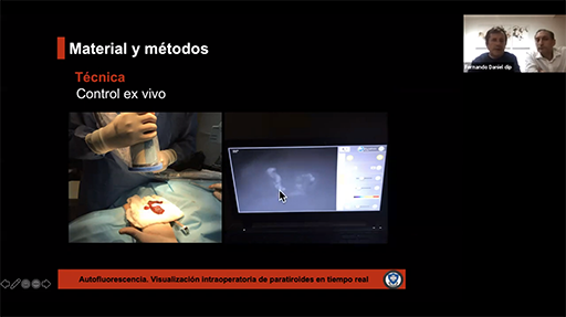
Indocyanine green (ICG) fluorescence imaging is increasingly used during thyroidectomy to help identify and preserve parathyroid glands and assess tissue perfusion. Using near-infrared fluorescence imaging, surgeons can visualize parathyroid vascularization in real time, supporting safer thyroid surgery and reducing the risk of postoperative hypoparathyroidism.
This dosing and timing chart provides guidance on recommended ICG administration for thyroidectomy procedures, including intravenous injection protocols, dosage considerations, and expected fluorescence detection timing to optimize fluorescence imaging during thyroid surgery.
Sección:
Title: Dosing & Timing Chart, Tabla:
Columna1:
Purpose:
, Contenido:
Visualization of parathyroid glands
Columna1:
Injection Type:
, Contenido:
Intravenous
Columna1:
Dilution (25 mg in 10 mL of sterile water - 2.5mg/mL):
, Contenido:
Yes
Columna1:
Requires Flush with Sterile Water:
, Contenido:
Yes
Columna1:
Proposed Dosage:
, Contenido:
1 mL
Columna1:
Injection Time:
, Contenido:
After thyroid gland dissection
Columna1:
First Indocyanine Green Detection:
, Contenido:
30–60 seconds after administration
Columna1:
Indocyanine Green Duration:
, Contenido:
60 seconds–3 minutes
Columna1:
Camera Requirements (handheld device, laparoscope or both):
, Contenido:
Handheld
Columna1:
Tips & Tricks:
, Contenido:
Check perfusion of parathyroid glands at the time of assessment.
Title: Dosing & Timing Chart, Contenido: , Tabla:
Columna1:
Purpose:
, Contenido:
Guided dissection during thyroid gland exposure
Columna1:
Injection Type:
, Contenido:
Intravenous
Columna1:
Dilution (25 mg in 10 mL of sterile water - 2.5mg/mL):
, Contenido:
Yes
Columna1:
Requires Flush with Sterile Water:
, Contenido:
Yes
Columna1:
Proposed Dosage:
, Contenido:
1 mL
Columna1:
Injection Time:
, Contenido:
During thyroid gland dissection
Columna1:
First Indocyanine Green Detection:
, Contenido:
30–60 seconds after administration
Columna1:
Indocyanine Green Duration:
, Contenido:
60 seconds–3 minutes
Columna1:
Camera Requirements (handheld device, laparoscope or both):
, Contenido:
Handheld
Columna1:
Tips & Tricks:
, Contenido:
Check vasculature surrounding gland during superior pole dissection
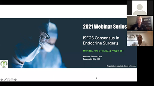
Descripción: In 2019, The International Society for Fluorescence Guided Surgery hosted a series of Delphi surveys meant to standardize the use of ICG in various specialties.
During this webinar, Dr. Michael Bouvet and Dr. Fernando Dip discuss ICG and fluorescence as related to endocrine surgery.
Consensus in Endocrine Surgery
Michael Bouvet and Fernando Dip
In 2019, The International Society for Fluorescence Guided Surgery hosted a series of Delphi surveys meant to standardize the use of ICG in various specialties.
During this webinar, Dr. Michael Bouvet and Dr. Fernando Dip discuss ICG and fluorescence as related to endocrine surgery.
25-06-2019