Título del video: Ultra-Low Dose ICG: Eliminating Hepatic Background Fluorescence for Consistent Biliary Visualization in All Cholecystectomy Scenarios
Título del video: Indocyanine Green Fluorescence-Guided Cholecystectomy and Biliary Safety in Acute Cholecystitis: A Systematic Review and Meta-Analysis
Descripción: Nuestro invitado, Enrique Lanzarini, habla sobre el rol de la cirugía guiada por fluorescencia en el tratamiento del cáncer gástrico, abordando cómo esta tecnología contribuye a una mejor visualización intraoperatoria y a una toma de decisiones más precisa.
Indocyanine green (ICG) fluorescence imaging is used in laparoscopic sleeve gastrectomy to evaluate gastric perfusion and support intraoperative decision-making. By providing real-time visualization of blood supply, this technique helps surgeons identify critical vascular structures and optimize staple line integrity.
In the EMEA region, standardized dosing and timing protocols aim to improve consistency in fluorescence imaging and enhance surgical safety. Although its routine use in bariatric surgery remains under evaluation, ICG has shown potential in assessing tissue perfusion and reducing ischemic complications.
Sección:
Tabla:
Columna1:
Region of interest
, Contenido:
Proximal stomach
Columna1:
Dose
, Contenido:
5 -15 mg bolus
Columna1:
Route
, Contenido:
I.V.*
Columna1:
Injection time
, Contenido:
Intraoperatively
Columna1:
First ICG detection
, Contenido:
30-60 sec
Columna1:
ICG Duration
, Contenido:
3-4 min
Columna1:
Camera requirements
, Contenido:
Laparoscope, NIR light source camera
Columna1:
References (full ref details at end of this guide doc)
Indocyanine green (ICG) fluorescence imaging is increasingly used in oesophagectomy to assess gastric conduit perfusion and guide anastomotic site selection. Adequate vascularization is critical, as poor perfusion is a major factor in anastomotic leak, one of the most serious postoperative complications.
In the EMEA region, standardized dosing and timing protocols help ensure consistent fluorescence imaging, supporting intraoperative decision-making and potentially reducing perfusion-related complications.
Sección:
Tabla:
Columna1:
Region of interest
, Contenido:
Gastric conduit
Columna1:
Dose
, Contenido:
5 mg bolus
Columna1:
Route
, Contenido:
I.V.*
Columna1:
Injection time
, Contenido:
Intraoperatively
Columna1:
First ICG detection
, Contenido:
60 – 100 sec
Columna1:
ICG Duration
, Contenido:
3-4 min
Columna1:
Camera requirements
, Contenido:
Laparoscope, NIR light source camera
Columna1:
References (full ref details at end of this guide doc)
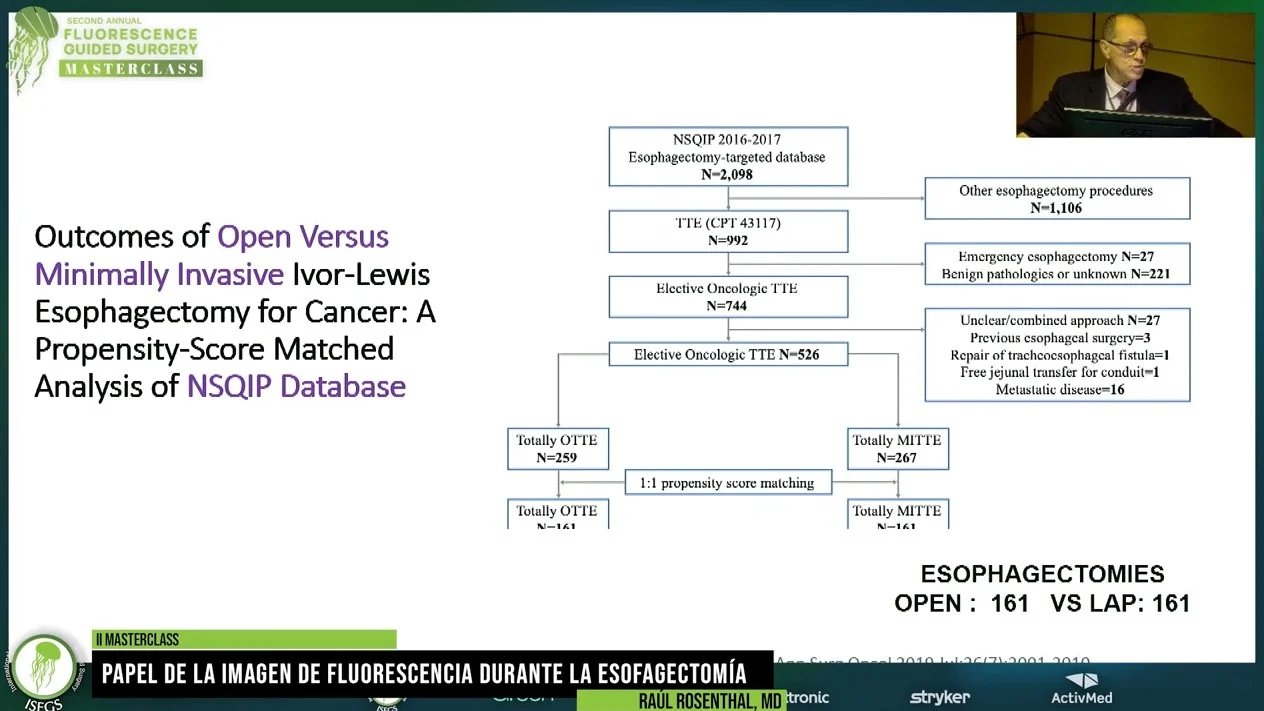
Indocyanine green (ICG) fluorescence imaging is increasingly used during esophagectomy to assess gastric conduit perfusion and support safe esophagogastric anastomosis. Near-infrared fluorescence angiography allows surgeons to visualize real-time blood flow to the gastric conduit, helping identify well-perfused tissue and reduce the risk of anastomotic complications.
This dosing and timing chart outlines recommended ICG administration for esophagectomy procedures, including intravenous injection protocols, dosage guidance, and expected fluorescence detection times to optimize intraoperative perfusion assessment during fluorescence-guided surgery.
Sección:
Title: Dosing & Timing Chart, Tabla:
Columna1:
Purpose:
, Contenido:
Perfusion assessment
Columna1:
Injection Type:
, Contenido:
Intravenous
Columna1:
Dilution (25 mg in 10 mL of sterile water - 2.5mg/mL):
, Contenido:
Yes
Columna1:
Requires Flush with Sterile Water:
, Contenido:
Yes
Columna1:
Proposed Dosage:
, Contenido:
3 mL
Columna1:
Injection Time:
, Contenido:
Intraoperatively
Columna1:
First Indocyanine Green Detection:
, Contenido:
30–60 seconds after administration
Columna1:
Indocyanine Green Duration:
, Contenido:
60 seconds–3 minutes
Columna1:
Camera Requirements (handheld device, laparoscope or both):
, Contenido:
Both
Columna1:
Tips & Tricks:
, Contenido:
Perform tissue vitality assessment, as necessary, prior to resection. Repeat assessment after anastomosis.