Título del video: Development and Preclinical Validation of Hepatocyte Growth Factor (c-Met) Peptide for Targeted Fluorescence Near-Infrared Imaging in Human Lung Adenocarcinoma
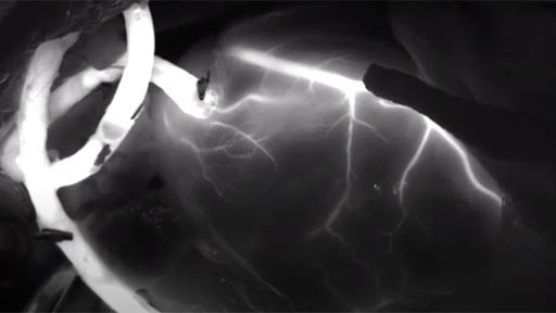
Título del video: NIRF-Guided Coronary Artery Bypass Grafting (CABG)
Speakers: Roderick Peul, MD
Descripción: This video demonstrates the use of near-infrared fluorescence (NIRF) imaging during coronary artery bypass grafting. The technique allows real-time assessment of graft perfusion and patency, supporting intraoperative decision-making and improving surgical outcomes.
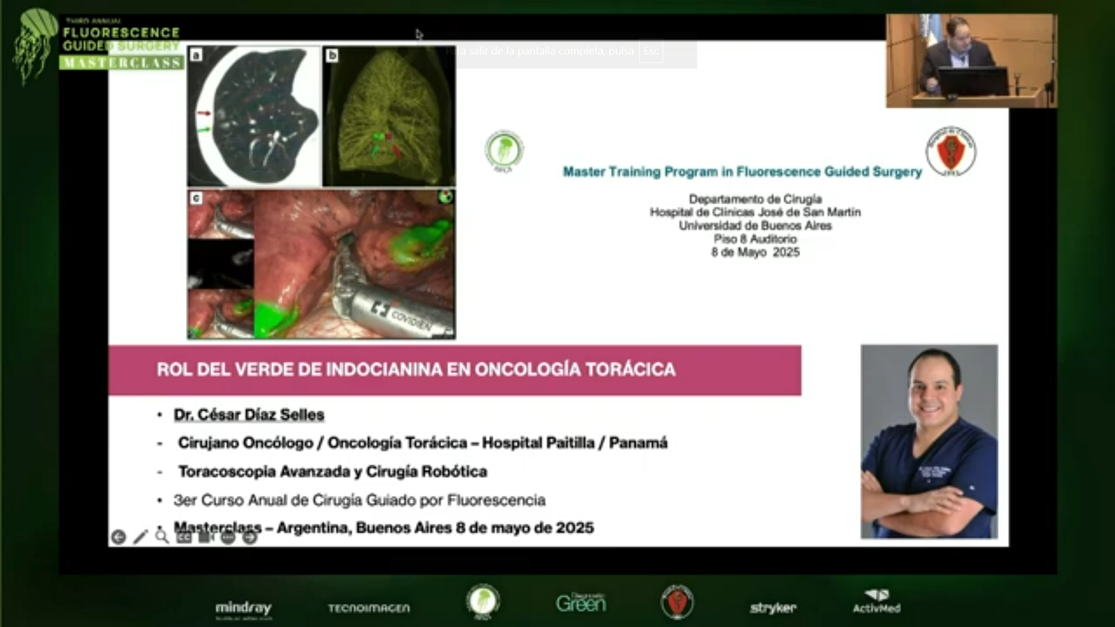
Indocyanine green (ICG) fluorescence imaging is increasingly used in thoracoscopic segmentectomy (VATS) to identify intersegmental planes, localize pulmonary nodules, and assess surgical margins. Precise delineation of segmental anatomy is critical in lung-sparing procedures, and ICG enables real-time visualization without the limitations of conventional inflation techniques.
ICG-based near-infrared imaging has demonstrated high accuracy in defining anatomical boundaries and improving intraoperative navigation, supporting more precise and minimally invasive lung resections.
In the EMEA region, standardized dosing and timing protocols help ensure consistent fluorescence imaging and enhance surgical precision in thoracic procedures.
Sección:
Tabla:
Columna1:
Region of interest
, Contenido:
Verification of anatomic segment borders
Columna1:
Dose
, Contenido:
0.15 mg/kg
Columna1:
Route
, Contenido:
I.V.*
Columna1:
Injection time
, Contenido:
Intra-operatively
Columna1:
First ICG detection
, Contenido:
10-25sec after peripheral injection
Columna1:
ICG Duration
, Contenido:
30-120 sec
Columna1:
Camera requirements
, Contenido:
Laparoscope, NIR light source camera
Columna1:
References (full ref details at end of this guide doc)
Título del video: Intraoperative fluorescent Imaging during CABG surgery
Speakers: Derek Muehrcke
Descripción: Coronary Artery Bypass grafts are the first step during Cardiac surgery. This video demonstrates every step leading up to the surgery and how ICG is used to help to detect any unexpected complications within the first minute and a half of surgery. Once these complications are rectified the patient shows improved outcomes and their hospital costs are lowered a shortened recovery time.
Intraoperative fluorescent Imaging during CABG surgery
Coronary Artery Bypass grafts are the first step during Cardiac surgery. This video demonstrates every step leading up to the surgery and how ICG is used to help to detect any unexpected complications within the first minute and a half of surgery. Once these complications are rectified the patient shows improved outcomes and their hospital costs are lowered a shortened recovery time.
Abstract
Background
Anastomotic leakage in esophagectomy is a serious complication, and assessing blood perfusion in the conduit can help minimize this risk. Indocyanine green is the most widely used method to assess tissue blood flow; however, this technique has disadvantages. Evaluating tissue oxygen saturation in the gastric conduit during thoracic esophagectomy compared with indocyanine green blood perfusion assessment addresses these disadvantages and can be performed easily and repeatedly.
Methods
This was a prospective study of patients with esophageal cancer...
We use cookies and similar technologies to improve your experience on our website.